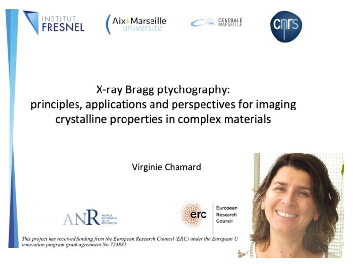
VIDEO - CoWork series - X-ray Bragg ptychography: principles, applications and perspectives for imaging crystalline properties in complex materials, with Virginie Chamard


VIDEO - CoWork series - X-ray Bragg ptychography: principles, applications and perspectives for imaging crystalline properties in complex materials, with Virginie Chamard
In this presentation, I will first review the principles of the Bragg ptychography approach, before detailing a selection of scientific results obtained with this novel crystalline microscopy. I will finally discuss some exciting perspectives opened by the advent of the fourth generation synchrotron sources.
Speaker: Virginie Chamard, Institute Fresnel Marseille, France
The webinar is part of the LINXS webinar series, CoWork. The CoWork webinar series is dedicated to the exploitation of the coherence properties of X-rays for advanced materials characterization, with a special focus on inverse microscopy techniques, such as Coherent Diffraction Imaging (CDI), Ptychography and Holography.
Abstract
3D X-ray Bragg ptychography has been proposed as a mean to map crystalline properties, in 3D with a high spatial resolution (a few tens of nm) and a high strain sensitivity (a few 10-4) [1], able to fill the experimental gap, which results from requirements imposed by other approaches. This encompasses 3D materials, which cannot be thinned down to meet electron microscopy constraint and/or which present large displacement fields and/or with dimension much larger than the accessible x-ray coherence volume.
Since the first demonstration of Bragg ptychography, specific efforts have been made on the methodological developments to ease its use [2-4]. In parallel, a series of novel results have been obtained based on the investigations of complex crystalline structures. As an illustration, one can cite the 3D strain mapping in semi-conductor-based nanostructures [3], the visualization of antiphase domain boundaries in metallic alloy [5] or the description of coherent crystalline domains in biominerals [6], to cite a few.
In this presentation, I will first review the principles of the Bragg ptychography approach, before detailing a selection of scientific results obtained with this novel crystalline microscopy. I will finally discuss some exciting perspectives opened by the advent of the fourth generation synchrotron sources.
[1] Godard et al., Nature Commun. (2011).
[2] Godard et al., Optics Express (2012).
[3] Hruszkewycz et al., Nature Materials (2017)].
[4] Hill et al., Nanoletters (2018).
[5] Kim et al., Phys. Rev. Letters (2018).
[6] Mastropietro et al., Nature Materials (2017).
Biography
V. Chamard is CNRS appointed x-ray scattering physicist, with strong expertise in synchrotron-based nanosciences. After her first work in nano-material science, she joined Institut Fresnel in 2009, a well-recognized institute in the fields of electromagnetism, photonics and signal processing. Within this exciting scientific environment, she established novel x-ray microscopy approaches, based on state-of-the art super-resolution microscopy and inversion algorithms, among which Bragg ptychography microscopy. Her group is gathering expertise in inverse problems and coherent (x-ray and optical) microscopy, a rather unique configuration worldwide. The originality of her developments is to combine extended methodological analysis and applications to complex materials, including semiconductor based devices, metallic alloy and biominerals. Her current research project aims at understanding the physico-chemical processes at play during biomineralization, using state-of-the art optical and x-ray microscopy approaches.

