Workshop on 3D histology offered support for researchers interested in synchrotron radiation imaging of soft tissue
Partcipants pictured at LINXS at the hackathon which folllowed on the workshop. It was targeted at researchers who wanted support with analysing their already acquired data.
In December 2023, the Biomedical Imaging working group organised a workshop on 3D histology with synchrotron micro-tomography. The aim was to offer an appetiser for people who are doing imaging of soft tissue but do not have any experience of synchrotron radiation. More than 40 people attended the event, which was completely full, from the Lund University Medical Faculty, MAX IV, and other places in Sweden and abroad.
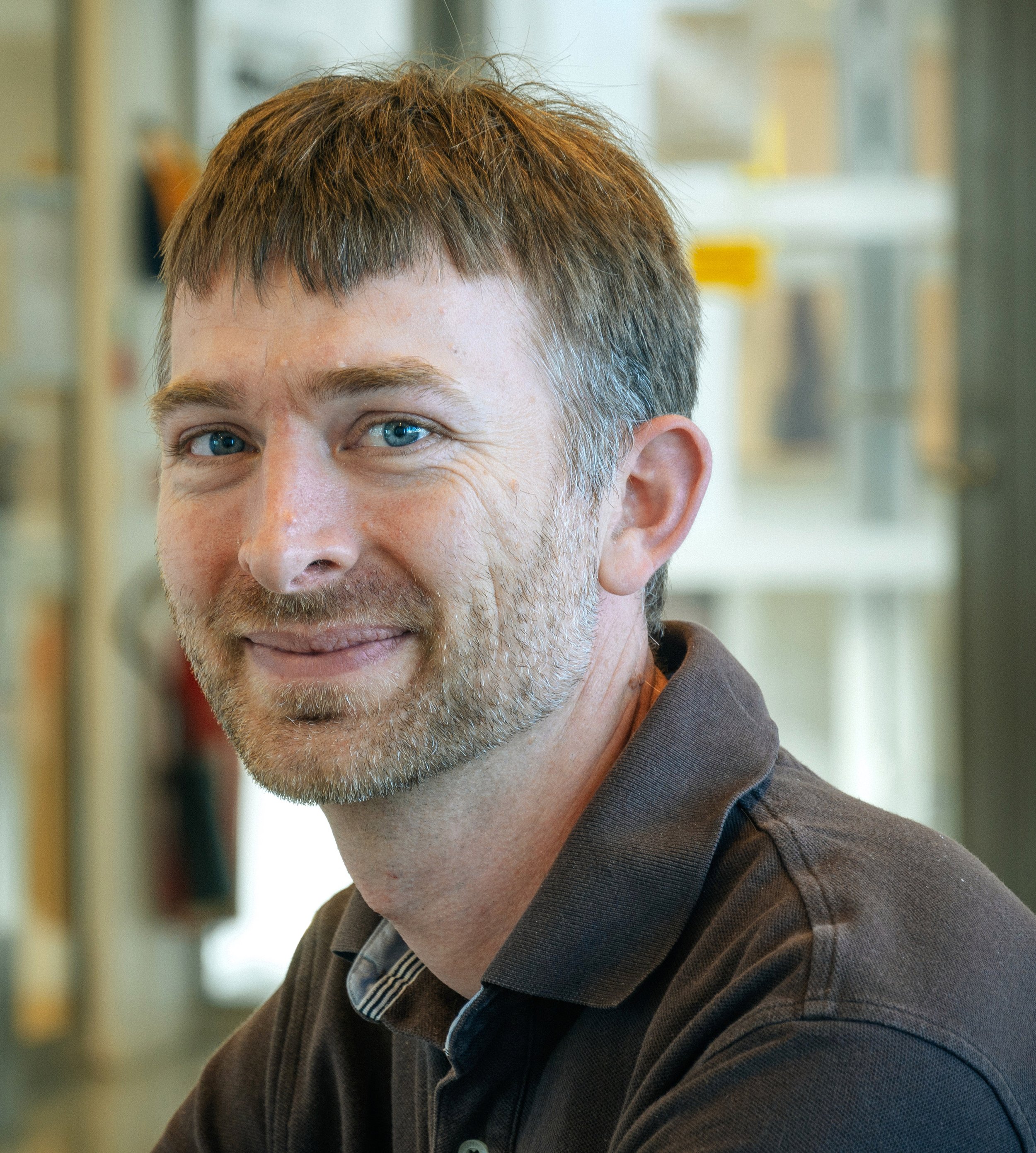
Martin Bech is leader of the Biomedical Imaging working group, and a senior lecturer at the division of Medical Radiation Physics, Clinical Sciences, Lund University.
The programme included presentations by scientists and experts who gave examples of how they have used and are using 3D-imaging methods, virtual histology and synchrotron based micro-tomography to image soft tissue, ranging from bones to hearts. There was also time for discussions on how one can get access to beamlines and large-scale infrastructures, and how to extract, segment and analyse data from beamline experiments.
– It is all about breaking down initial barriers, says Martin Bech, who was one of the event organisers. He is leader of the Biomedical Imaging working group, and a senior lecturer at the division of Medical Radiation Physics, Clinical Sciences, Lund University.
He says that researchers often get alarmed by the long and tedious application procedure and may refrain from applying for beamtime.
– With this event, we tried to make it easier by highlighting how one can go about preparing for beamtime. By inviting experts and scheduling time for discussions, we tried to stimulate participants with no or little experience with synchrotron radiation to build networks with staff from different beamlines.
Hackathon followed - provided support for researchers to analyse data
The symposium was followed by a hackathon the next day, for researchers who wanted support with analysing their already acquired data. During the hackathon, the focus was on how to use commercial software for visualisation, and how to make use of AI to segment and analyse soft tissue imaging data. In the afternoon, participants worked independently, with support from data analysis experts.
– We got a lot of positive feedback on the hackathon as well as on the symposium. I think people enjoyed that we gave a lot of practical tips along with inspiring examples, says Martin Bech.
Further reflecting on the two events, he notes that he was surprised by the fact that even if people are working on different samples, and are from different disciplines, they often have similar challenges, and find inspiration in the same presentations.
– By showing that different problems can be solved with common imaging tools, our working group can offer a platform for broader knowledge and understanding of ways to approach for example soft tissue imaging. It also encourages knowledge exchange amongst researchers with different backgrounds.
He emphasises that the overall aim of the working group is to facilitate current and future imaging at MAX IV and ESS for bio-medical research and development, with focus on methods and tools to improve data analysis and segmentation.
– We need to get researchers and data experts together to see how we can improve what is already out there and come up with new ways of working. This motivation is what lies at the heart of the working group and also my own work. As a physicist I want to develop tools that can support researchers in other fields as well.
Read more about the theme and the working group


